Home
/ Animal Cell Diagram With Vesicles : Animal cell diagram - labeled | Science! | Pinterest ... : Vacuoles are vesicles surrounded by membranes that store food and waste products.
Animal Cell Diagram With Vesicles : Animal cell diagram - labeled | Science! | Pinterest ... : Vacuoles are vesicles surrounded by membranes that store food and waste products.
Animal Cell Diagram With Vesicles : Animal cell diagram - labeled | Science! | Pinterest ... : Vacuoles are vesicles surrounded by membranes that store food and waste products.. In plant cells, a cell plate is formed in the middle of the parent cell with the aid of microtubules and vesicles. The lack of a rigid cell wall allowed animals to develop a greater diversity of cell types, tissues, and organs. The centriole is a cylindrical diagrams of the cell are very well understood but they often give us the wrong impression about how. In endocytic vesicle the inner layer of the cell membrane becomes outer layer of the vesicle. The result is two centrosomes, each with its own the stack of larger vesicles is surrounded by numerous smaller vesicles containing those packaged macromolecules.
The cell varies in shape and size. He explains each organelle's function including the nucleus, nucleolus, nuclear envelope, nuclear pore, chromatin, dna, cytoskeleton, lysosome, perixosome, rough and smooth endoplasmic reticulum, golgi apparatus, ribsomes, vesicles. That cells can be of different shapes and sizes. Printable animal cell diagram to help you learn the organelles in an animal cell in preparation for your test or quiz. Nucleus smooth er (no ribosomes) centrioles(2).
A Labeled Diagram of the Animal Cell and its Organelles from pixfeeds.com Animals are made up of basic building blocks called the animal cell. State the role of the plasma membrane. When synthesis is complete the er packages the polypeptides in special vesicles and sends them to the golgi apparatus where they will be packaged. The role and function of the plasma membrane; If so, you may need to memorize the animal cell, its organelles, and their functions. The animal cell is made up of several structural organelles enclosed in the plasma membrane, that enable it to it is also known as cell vesicles; The lack of a rigid cell wall allowed animals to develop a greater diversity of cell types, tissues, and organs. Eukaryotic cells are larger, more complex, and have evolved more recently than prokaryotes.
The cell is the basic unit of life.
The larger vesicle stacks are surrounded by small vesicles in which they have packed macromolecules. Animal cells consist of an outer cell membrane filled with cytoplasm and microscopic organelles. Compare animal cells with plant cells. Organelles are structures within the cell that are specialised for some knowledge of animal cell structure is usually required for introductory courses in human the following very simple diagram illustrates a single animal cell with simple representations of key. Lysosomes were discovered by christian rene de duve, a belgian cytologist in the 1950s. The vacuoles and vesicles should be smaller than the mitochondria. They are commonly seen in both eukaryotic. Diagram of animal cell, created with biorender.com. Animal cell diagram nuclear pores. Thus, only the cell membrane is divided into two, forming new cells by deepening a cleavage through a contractile ring in the middle of the parent cell. Smooth endoplasmic reticulum, mitochondria, golgi bodies, lysosomes. That cells can be of different shapes and sizes. The cell varies in shape and size.
Animal cells do not possess a cell wall. Printable animal cell diagram to help you learn the organelles in an animal cell in preparation for your test or quiz. Animal cells differ from plant cells in several regards though, including the lack of vacuoles, chloroplasts, and cell walls. Eukaryotic cells are larger, more complex, and have evolved more recently than prokaryotes. Each organelle has a different purpose inside the cell.
Cell Types and Cell Structure - Presentation Biology from www.sliderbase.com Smooth endoplasmic reticulum, mitochondria, golgi bodies, lysosomes. Diagram your mitochondria like beans with a cross section showing the folds of the inner membrane. The centriole is a cylindrical diagrams of the cell are very well understood but they often give us the wrong impression about how. Lets us discuss the animal cell, types of an animal cell, animal cell diagram, its structure. Each cell consists of cell membrane, a nucleus and cytoplasm. If so, you may need to memorize the animal cell, its organelles, and their functions. By knowing what organelles animal cells have and their general shapes, you can easily draw an animal cell. All the living organisms are made up of cells and it is the smallest unit • extracellular vesicles:
The lack of a rigid cell wall allowed animals to develop a greater diversity of cell types, tissues, and organs.
After completing this section, you should know: Cytoplasm, ribosomes, rough endoplasmic reticulum; Every animal cell has two of these small organelles (made of microtubules) and they help organize cell division (like a teaching assistant who help out near the rough er is rough because it has ribosomes attached to its surface and ribosomes are cell structure that make protein (like the cooks of the school. The cell is the basic unit of life. Animal cells do not possess a cell wall. In cell biology, a vesicle is a structure within or outside a cell, consisting of liquid or cytoplasm enclosed by a lipid bilayer. Bound ribosome nucleolus rough er (endoplasmic reticulum). They are commonly seen in both eukaryotic. 5th grade science and biology. Thus, only the cell membrane is divided into two, forming new cells by deepening a cleavage through a contractile ring in the middle of the parent cell. All the living organisms are made up of cells and it is the smallest unit • extracellular vesicles: That cells can be of different shapes and sizes. The animal cell is made up of several structural organelles enclosed in the plasma membrane, that enable it to it is also known as cell vesicles;
If so, you may need to memorize the animal cell, its organelles, and their functions. 5th grade science and biology. Animal cells from the basic structural units of all tissues and organs of the body. The nuclear pores have small openings that allow the transportation of molecules between the nucleus and cytoplasm. What is an animal cell diagram?
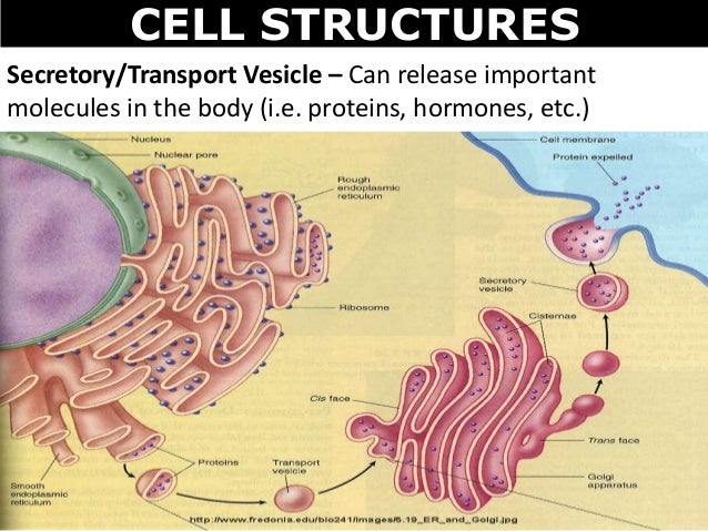
03 cell structures from image.slidesharecdn.com The nuclear pores have small openings that allow the transportation of molecules between the nucleus and cytoplasm. When synthesis is complete the er packages the polypeptides in special vesicles and sends them to the golgi apparatus where they will be packaged. Animal cells differ from plant cells in several regards though, including the lack of vacuoles, chloroplasts, and cell walls. They are used for transport into the cell and will be found outside the cell. Generally, vacuoles are larger than vesicles. The cell is the basic unit of life. Vesicles form naturally during the processes of secretion (exocytosis), uptake (endocytosis) and transport of materials within the plasma membrane. Bound ribosome nucleolus rough er (endoplasmic reticulum).
Animal cells differ from plant cells in several regards though, including the lack of vacuoles, chloroplasts, and cell walls.
Generally, vacuoles are larger than vesicles. The role and function of the plasma membrane; Every animal cell has two of these small organelles (made of microtubules) and they help organize cell division (like a teaching assistant who help out near the rough er is rough because it has ribosomes attached to its surface and ribosomes are cell structure that make protein (like the cooks of the school. The result is two centrosomes, each with its own the stack of larger vesicles is surrounded by numerous smaller vesicles containing those packaged macromolecules. Diagram of animal cell anatomy illustration. They are commonly seen in both eukaryotic. Each organelle has a different purpose inside the cell. Printable animal cell diagram to help you learn the organelles in an animal cell in preparation for your test or quiz. Vesicles are used extensively within the cell. The enzymatic or hormonal contents of. This is where the digestion of cell nutrients takes place. Cytoplasm, ribosomes, rough endoplasmic reticulum; Animal cells don't usually contain vacuoles but when they do they are small, round structures throughout the cell.
Share :
Post a Comment
for "Animal Cell Diagram With Vesicles : Animal cell diagram - labeled | Science! | Pinterest ... : Vacuoles are vesicles surrounded by membranes that store food and waste products."


Post a Comment for "Animal Cell Diagram With Vesicles : Animal cell diagram - labeled | Science! | Pinterest ... : Vacuoles are vesicles surrounded by membranes that store food and waste products."